In laparoscopic or robotic myomectomy, minimally invasive procedures, your surgeon accesses and removes fibroids through several small abdominal incisions.
Laparoscopic myomectomy. Your surgeon makes a small incision in or near your bellybutton. Then he or she inserts a laparoscope ― a narrow tube fitted with a camera ― into your abdomen. Your surgeon performs the surgery with instruments inserted through other small incisions in your abdominal wall.
Robotic myomectomy. Instruments are inserted through small incisions similar to those in a laparoscopic myomectomy, and the surgeon controls movement of instruments from a separate console.
Sometimes, the fibroid is cut into pieces and removed through a small incision in the abdominal wall. Other times the fibroid is removed through a bigger incision in your abdomen so it can be removed without being cut into pieces. Rarely, the fibroid may be removed through an incision in your vagina (colpotomy).
Laparoscopic and robotic surgery use smaller incisions than a myomectomy, or laparotomy, does. This means you may have less pain, lose less blood and return to normal activities more quickly than with a laparotomy.
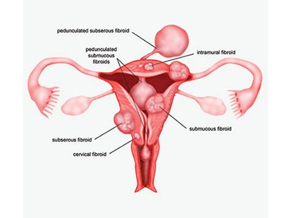
To treat fibroids that bulge significantly into your uterine cavity (submucosal fibroids), your surgeon may suggest a hysteroscopic myomectomy. Your surgeon accesses and removes fibroids using instruments inserted through your vagina and cervix into your uterus.
A hysteroscopic myomectomy generally follows this process:
Rarely, your surgeon may use a laparoscope inserted through a small incision in your abdomen to view the pelvic organs and monitor the outside of the uterus during a complicated hysteroscopic myomectomy.
At discharge from the hospital, your doctor prescribes oral pain medication, tells you how to care for yourself, and discusses restrictions on your diet and activities. You can expect some vaginal spotting or staining for a few days up to six weeks, depending on the type of procedure you've had.